
Surgical Magnification: A Comprehensive Guide to Loupes, Microscopes & Enhanced Precision
Imagine the most intricate task you’ve ever performed. Now, imagine doing it through a foggy window, with limited light, on a moving target where a millimeter’s error carries significant consequences. This is the inherent challenge of surgery with the naked eye. Human vision, remarkable as it is, has physiological limits in resolution, depth perception, and stamina under the demanding conditions of the operating room. So, what if you could operate with superhuman sight? What if you could see tissue planes with crystal clarity, distinguish micro-vessels with ease, and work with a precision that transcends natural ability?
This is not science fiction; it is the daily reality enabled by surgical magnification. Far from being a luxury or a niche tool for a few super-specialists, magnification has become a fundamental pillar of modern surgical practice. It is a critical investment that drastically improves clinical outcomes, reduces surgeon fatigue and career-limiting strain, and is the very enabler of minimally invasive techniques that define contemporary patient care. This comprehensive guide is designed for surgeons, surgical residents, and operating room professionals seeking authoritative, experience-based information. We will demystify the optical technology, explore the robust evidence behind its benefits, provide a detailed framework for selecting the right system, and outline the best practices for integration into your surgical workflow. Our goal is to empower you with the knowledge to make an informed decision that enhances your capability, protects your well-being, and elevates the standard of care you provide.
What is Surgical Magnification and Why is it Essential?
Beyond the Naked Eye: Defining the Technology
At its core, surgical magnification is any optical system designed to enlarge the surgeon’s view of the operative field. However, to dismiss it as merely “making things bigger” is to miss its profound sophistication. True surgical magnification systems are engineered around three interdependent optical principles critical for practical use:
- Working Distance: This is the optimal space between the surgeon’s eyes (or the front lens) and the surgical site. It is not a random measurement but a carefully chosen distance that allows for instrument manipulation and a comfortable, upright posture. An incorrect working distance forces the surgeon into a debilitating, hunched position.
- Depth of Field: This refers to the three-dimensional “slice” of space that remains in sharp focus at a given setting. A larger depth of field allows the surgeon to see a wound cavity in focus without constant re-adjustment, which is crucial for efficiency and spatial awareness.
- Resolution: This is the system’s ability to distinguish two closely spaced objects as separate. High resolution is what turns a blurry, enlarged image into a detailed, usable one, revealing subtle structures like nerve fibers or vessel walls.
The synergy of these three elements—customized to the surgeon’s anatomy and surgical specialty—is what transforms a simple magnifying glass into a precision surgical instrument.
The Proven Clinical Benefits: Evidence-Based Outcomes
The adoption of magnification is driven by a compelling body of clinical evidence and experiential reporting that underscores its necessity.
- Enhanced Precision & Accuracy: Magnification provides visual feedback that is otherwise impossible. Studies across specialties consistently demonstrate its impact. In microsurgery and plastic surgery, it is directly linked to higher patency rates in vascular anastomoses. In dentistry, it improves the detection of canal orifices and marginal integrity of restorations. In general surgery, it allows for more precise dissection in delicate areas like the biliary tree or during vascular procedures, reducing iatrogenic injury.
- Reduced Surgeon Fatigue: Ergonomics in surgery is a growing imperative. Magnification systems, when properly fitted, promote a neutral spine and head position. By bringing the target into clear focus without requiring the surgeon to hunch forward, they significantly reduce strain on the cervical and lumbar spine. Furthermore, reducing the constant muscular effort of visual accommodation (the eye’s focus mechanism) alleviates eye strain and headaches, allowing for sustained concentration during long procedures.
- Improved Patient Outcomes: The chain of logic is direct: enhanced precision leads to more accurate tissue handling, gentler dissection, and more meticulous repair. This translates to tangible patient benefits, including shorter operative times, lower rates of post-operative complications (e.g., leaks, infections, nerve damage), and improved healing potential. In fields like ophthalmology or nerve repair, it is not an enhancement but an absolute requirement for a successful outcome.
- Facilitation of Minimally Invasive Surgery: The entire philosophy of minimally invasive surgery (MIS) relies on working through small portals. Magnification, particularly Loupes, is what makes this feasible. It allows the surgeon to maintain a panoramic, magnified view of deep structures through a limited incision, making procedures like endoscopic carpal tunnel release or laparoscopic microsurgical techniques not only possible but routine.
Types of Surgical Magnification Systems
Surgical Loupes: The Surgeon’s Mobile Magnifier
Loupes are the most personal and ubiquitous form of magnification, offering a portable, head-mounted solution. They fall into two main optical categories:
- Galilean Systems: These use a simple convex objective lens and a concave eyepiece lens. They are lightweight, affordable, and typically offer lower magnification (2.0x to 3.5x) with a relatively larger field of view and good depth of field. They are an excellent, lower-cost entry point for procedures requiring moderate enlargement.
- Prismatic (Keplerian) Systems: These employ a series of prisms to fold the light path, allowing for a much longer optical path in a compact housing. This design supports higher magnification powers (3.5x to 8x and beyond) with superior optical clarity, resolution, and a longer working distance. The trade-offs are increased weight and higher cost. They are considered essential for specialties demanding high precision, such as periodontics, microsurgery, and intricate reconstructive work.
Loupes also come in two mounting styles:
* Through-the-Lens (TTL): Custom-built to the surgeon’s precise pupillary distance, vertex distance, and declination angle. They offer a perfect, seamless fit and optimized optical axis but cannot be adjusted or shared.
* Flip-Up: Feature adjustable eyepieces and mount on a frame, allowing them to be flipped up when not in use. They offer flexibility for sharing among users or for surgeons who wear glasses, but may be slightly bulkier and can lose alignment if adjusted frequently.
Operating Microscopes: The Gold Standard for High-Power Magnification
For procedures requiring extreme magnification and stability, the operating microscope is indispensable. It is a floor- or ceiling-mounted unit that provides a stereoscopic (3D) view at high powers (often 5x to 40x). Key features include:
* Variable Magnification: Hand-controlled or foot-pedal zoom and focus.
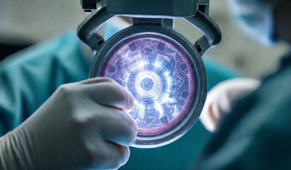
* Coaxial Illumination: A brilliant, shadow-free light source is directed down the same optical path as the view.
* Stereo Vision: Provides critical depth perception at high magnification.
* Assistant Scopes: Allow a second surgeon to share the identical visual field.
* Digital Integration: Built-in cameras for recording, broadcasting, and integration with surgical navigation or augmented reality systems. They are the cornerstone of neurosurgery, otology, vitreoretinal surgery, and microvascular reconstruction.
Headlights and Illumination Systems: The Crucial Companion
Magnification is fundamentally useless without adequate light. As magnification increases, the amount of light reaching the surgeon’s eyes decreases exponentially. A dedicated surgical headlight is therefore not an accessory but a core component. Modern LED systems offer bright, white, cool illumination with long battery life. Light can be delivered via fiber-optic cables or through newer LED-emitter modules mounted directly on the loupes. The integration of a high-quality light source with your magnification system is non-negotiable for achieving a clear, well-lit, and shadow-reduced field.
How to Choose the Right Magnification System: A Buyer’s Guide
Assessing Your Surgical Specialty and Needs
Your specialty and most common procedures are the primary determinants.
* Dentistry/Periodontics: High-power prismatic loupes (4.5x-6x+) are standard for endodontics and perio surgery, often with longer working distances.
* Plastic/Reconstructive Surgery: A range is used. 2.5x-3.5x Galilean loupes for general procedures, 4.5x-6x prismatic for microsurgery (e.g., free flaps, nerve repair). Operating microscopes are used for super-microsurgery.
* Neurosurgery & Ophthalmology: Primarily the domain of the operating microscope, though loupes may be used for opening/closing.
* General/Vascular Surgery: 2.5x-3.5x loupes are increasingly common for open vascular anastomoses, biliary surgery, and complex hernia repairs, improving dissection safety and suture precision.
Key Technical Specifications to Evaluate
- Magnification Power: More is not always better. Higher power reduces the field of view and depth of field. Choose the lowest power that allows you to see the necessary detail comfortably. For most general procedures, 2.5x-3.5x is sufficient. Reserve higher powers for truly micro-level work.
- Working Distance: This is critical for ergonomics. Measure the distance from your eye to your typical surgical site while maintaining a neutral, upright posture (chin slightly tucked, back straight). Vendors will use this measurement to build or adjust your loupes.
- Field of View vs. Depth of Field: Understand the trade-off. A wider field of view gives more context but often at the expense of a shallower depth of field. Your choice should balance the need for panoramic awareness versus the need to see varying depths in focus.
- Weight and Ergonomics: A difference of ounces matters over a 4-hour case. Lighter systems reduce neck load. Consider the weight distribution of the frame, loupes, and headlight battery pack.
The Fitting Process and Vendor Selection
A professional, in-person fitting is non-negotiable. This is a medical device, not an online purchase. A good vendor will:
* Take precise anatomical measurements.
* Discuss your surgical practice in detail.
* Allow you to trial different models.
* Provide thorough training on use and maintenance.
Ask about warranty length, repair turnaround time, and lens coating options (e.g., anti-fog, anti-reflective). Budget for the total system (loupes, frame, light) and view it as a long-term investment in your career health and surgical capability.
Best Practices for Using and Maintaining Your Equipment
Achieving Optimal Ergonomics in the OR
- Posture First: Adjust the operating table height so that when you look through your loupes with a straight back, the target is in perfect focus. Do not bend to meet the loupes.
- Microbreaks: Incorporate brief moments to look away into the distance during long cases to relax your ciliary muscles.
- Strengthening: Regular exercises to strengthen core and postural muscles will support your endurance in the OR.
Daily Care, Cleaning, and Maintenance Protocols
- Cleaning: Use only lens-specific tissue or microfiber cloths. Apply lens cleaner to the cloth, not directly to the lens, to avoid seepage into housing. Gently wipe in a circular motion.
- Storage: Always place loupes in a hard, protective case. Never leave them on a surgical drape where they can be knocked off or sat on.
- Alignment Check: Periodically, look at a straight horizontal line (e.g., a door frame) with both eyes open. If you see two separate, misaligned lines, your loupes need professional servicing.
- Professional Servicing: Schedule an annual check-up with your vendor for deep cleaning, alignment verification, and hardware inspection.
Common Pitfalls and How to Avoid Them
- Incorrect Working Distance: Leads to hunched posture. Solution: Remeasure and refit if necessary.
- Poor Posture: Slouching to compensate for poor table height or incorrect loupes. Solution: Be militant about table height and your own posture.
- Using Damaged Equipment: Scratched lenses or misaligned optics cause eye strain and headaches. Solution: Immediate repair.
- Neglecting Illumination: Trying to “make do” with overhead OR lights. Solution: Invest in and use a dedicated headlight every time you use loupes.
Frequently Asked Questions (FAQ)
Q: At what stage in my surgical training should I invest in loupes?
A: Early adoption, typically during residency, is highly recommended. It allows you to develop fine motor skills under magnification from the start, building proper ergonomic habits that will protect you throughout your career. Learning to operate with magnification is a skill in itself.
Q: Are expensive prismatic loupes worth it over Galilean systems?
A: It depends entirely on your needs. If your specialty requires magnification above 3.5x, a longer working distance, or superior resolution for delicate structures (e.g., vessels <1mm), then prismatic loupes are worth the investment. For general procedures at lower magnifications, a high-quality Galilean system may be perfectly adequate and more budget-friendly.
Q: How often do surgical loupes need to be re-aligned or serviced?
A: With careful handling and proper storage, a well-made pair can go years without needing service. However, an annual professional check-up is wise. They must be serviced immediately if they are dropped or if you experience any visual discomfort, diplopia (double vision), or headache while using them.
Q: Can I use the same loupes for different procedures?
A: This is challenging because working distance is fixed. If your typical procedures (e.g., superficial skin surgery vs. deep cavity work) require significantly different working distances, one pair will force you into poor posture for one of them. Some surgeons do own multiple pairs for this reason.
Q: How does surgical magnification integrate with digital recording and augmented reality?
A: This is the frontier. Beam-splitters can be added to loupes and microscopes to feed a video camera for recording, telementoring, or live broadcasting. Augmented reality (AR) systems are emerging that can project pre-operative imaging (like CT scans) or navigation data directly into the surgeon’s visual field through the oculars, creating a hybrid real-time/augmented view.
Conclusion
Surgical magnification represents far more than an optical aid; it is a critical investment in a surgeon’s most vital assets: precision, ergonomic health, and career longevity. The evidence is clear that it enhances patient safety and outcomes by enabling a level of visual acuity and technical accuracy that the naked eye cannot achieve.
The final, most crucial piece of advice is this: prioritize the fitting and the education over the specifications on a brochure. The most expensive, highest-magnification loupes in the world are a liability if they are not perfectly tailored to you. This technology must become a seamless, comfortable extension of your own senses. Consult with experienced mentors about their equipment choices and lessons learned. Schedule professional fittings with multiple reputable vendors to feel the difference. Ground your decision in the peer-reviewed literature for your specific field.
Adopting and mastering surgical magnification, with proper training and a commitment to ergonomics, is a definitive step toward advancing both your personal practice and the broader standard of surgical care. It is a commitment to seeing—and therefore doing—better.
p>