
Headlamp Surgery: A Comprehensive Guide to This Minimally Invasive Procedure
Introduction
Imagine living with chronic sinus pain that medication can no longer touch, or persistent back pain that radiates down your leg, making every step a challenge. The traditional solution often involved a daunting prospect: major surgery with large incisions, significant pain, and a long, arduous recovery. For many patients, this fear can be a barrier to seeking the treatment they need.
Today, a revolution in surgical technique offers a different path. You may have heard the term “Une lampe frontale surgery” from a friend, in an online forum, or even from a doctor. While it sounds almost rudimentary, this colloquial term points to one of the most significant advances in modern medicine: the shift towards minimally invasive surgery (MIS) powered by enhanced visualization.
This guide is designed to demystify “headlamp surgery.” We will clarify that it is not a single procedure, but rather an approach and philosophy used across various surgical specialties. Our goal is to provide you with authoritative, clear, and patient-centered information—compiled from established surgical principles and medical literature—to help you understand what this approach entails, its remarkable benefits, and what you can realistically expect. Whether you are in the early stages of research, investigating specific procedures, or considering how to find a qualified surgeon, this post will address your core questions and empower you for informed conversations with your healthcare team.
What is Headlamp Surgery? Demystifying the Term
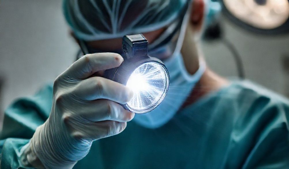
At its most literal, a headlamp is a light source worn on the surgeon’s forehead. However, the patient-coined term “headlamp surgery” symbolizes much more. It represents the entire ecosystem of technology that allows surgeons to operate with extraordinary precision through the smallest possible openings, minimizing disruption to the body.
Beyond the Literal Meaning
The headlamp itself is just one component. The true essence of “headlamp surgery” lies in the marriage of illumination et un fonction de grossissement. This combination transforms the surgical field, allowing a surgeon to see fine details and navigate complex anatomy that would be impossible to distinguish with the naked eye alone. It’s the hallmark of a minimally invasive mindset: achieving major surgical goals with minor access.
The Core Technology: Visualization and Precision
The “headlamp” metaphor covers several key tools:
- Surgeon’s Loupes: These are custom magnifying glasses (often 2.5x to 5.5x magnification) worn by the surgeon. They provide a stable, magnified, and illuminated view of the surgical site, essential for procedures requiring fine suturing or dissection in fields like plastic surgery, hand surgery, and some ENT and spine operations.
- Operating Microscopes: For the most delicate work, such as in neurosurgery or microvascular surgery, a high-powered operating microscope is used. This sophisticated tool provides intense, coaxial (shadow-free) light and much higher levels of magnification (often 10x to 40x), giving the surgeon a detailed, three-dimensional view of structures like nerves, blood vessels, and the spinal cord.
- Endoscopes and Rigid Scopes: This is where the technology creates entirely new pathways. An endoscope is a thin, tubular instrument with a high-resolution camera and a light at its tip. It is inserted through a natural opening (like the nostril) or a tiny incision. The camera transmits images to a high-definition monitor, allowing the surgeon to operate indirectly with specialized instruments. This is the cornerstone of procedures like endoscopic sinus surgery or arthroscopic knee surgery.
Common Surgical Fields Utilizing This Approach
The “headlamp surgery” approach is a cornerstone in several specialties:
- Otolaryngology (ENT): This is one of the most common areas. Chirurgie Sinusienne Endoscopique uses scopes through the nostrils to treat chronic sinusitis, remove polyps, or address skull base tumors without any external incisions. Pituitary tumor removal is also often performed this way.
- Neurosurgery: Delicate brain and nerve procedures rely heavily on this approach. Microvascular Decompression for trigeminal neuralgia uses a microscope through a small opening behind the ear to place a protective pad between a nerve and a blood vessel. Similarly, clipping a brain aneurysm requires the precision of microscopic visualization.
- Spine Surgery: This field has been transformed by minimally invasive techniques. A microdiscectomy uses a microscope or loupes to remove a herniated disc fragment through an incision often less than an inch long, relieving nerve pressure. Minimally Invasive Spinal Fusion uses specialized tubular retractors and visualization tools to place hardware and bone graft with minimal muscle damage.
Key Benefits of the Headlamp Surgery Approach
The advantages of this high-precision approach are substantial for both the patient and the surgeon, creating a win-win scenario that drives better overall outcomes.
For the Patient: Enhanced Recovery and Outcomes
This is where patients feel the most direct impact:
- Less Trauma, Less Pain: Smaller incisions mean less cutting of skin, muscle, and soft tissue. This directly translates to significantly less post-operative pain and a reduced need for strong pain medications.
- Minimal Scarring: Tiny incisions lead to tiny, often barely noticeable scars. In endoscopic approaches through natural openings, there are no visible scars at all.
- Reduced Risk Profile: Smaller wounds generally mean less blood loss during surgery and a lower risk of post-operative infection and bleeding complications.
- Faster Recovery: With less tissue disruption, the body can heal more quickly. This often results in shorter hospital stays (sometimes same-day discharge) and a faster return to daily activities, work, and hobbies.
- Potential for Better Long-Term Results: Enhanced visualization allows the surgeon to more accurately target the problem while preserving healthy surrounding structures. This precision can lead to more complete treatment of the condition and potentially better long-term functional outcomes.
For the Surgeon: Superior Visualization and Control
The benefits for the surgical team are what make these patient advantages possible:
- A Magnified, Illuminated “Roadmap”: The surgeon gains a crystal-clear view of delicate anatomical structures—tiny nerves, blood vessels, and membranes—that are critical to preserve.
- Navigation in Tight Spaces: The technology allows the surgeon to work effectively in confined anatomical corridors (like the nasal cavity or spinal canal) without needing to create a large access window.
- Improved Tissue Discrimination: The enhanced view helps in accurately distinguishing between diseased tissue (e.g., a tumor, herniated disc, or infected sinus tissue) and healthy tissue that should be spared, promoting more conservative and effective surgery.
What to Expect: Before, During, and After Procedure
Understanding the journey can alleviate anxiety and help you prepare effectively.
The Consultation and Pre-Operative Phase
This is the most critical planning stage.
* Thorough Diagnosis: Your surgeon will rely on detailed imaging—such as CT scans, MRI, or X-rays—to precisely map the anatomy of your condition and plan the surgical approach.
* Candidacy Discussion: Not everyone is an ideal candidate for a minimally invasive approach. Your surgeon will evaluate your specific anatomy, the nature and extent of your condition, your overall health, and previous surgeries to determine the best option for you.
* Verifying Surgeon Credentials: Do not hesitate to ask about your surgeon’s experience and training specifically in minimally invasive techniques. Ask if they have completed a fellowship in their subspecialty (e.g., Minimally Invasive Spine Surgery, Rhinology).
A Step-by-Step Look at the Surgical Day
- Anesthésie : La plupart de ces interventions sont réalisées sous anesthésie générale, meaning you will be completely asleep and pain-free.
- The Surgical Process: Once you are asleep, the team will position you carefully. The surgeon will then use the chosen visualization tool—loupes, microscope, or endoscope. For endoscopic or microscopic procedures, the surgeon’s focus will often be on a video monitor displaying the magnified surgical field. A skilled surgical assistant or nurse is vital in managing the technology and instruments.
- Team-Based Approach: Remember, this is a team effort involving the surgeon, anesthesiologist, surgical technicians, and nurses, all focused on your safety and the procedure’s success.
The Recovery Pathway and Post-Op Care
Recovery varies by procedure, but general principles apply:
* Hospital Stay: Many minimally invasive procedures are outpatient (same-day discharge). More complex surgeries may require a 1-3 day hospital stay.
* Gestion de la douleur : Pain is typically managed effectively with oral medications. The goal is comfort, not complete numbness.
* Activity & Follow-up: You will receive specific instructions on wound care, activity restrictions (like lifting limits or bending), and the timeline for returning to driving and work. Attending all follow-up appointments is crucial for monitoring your healing.
* Warning Signs: Your care team will instruct you on signs of potential complications to watch for, such as fever, excessive redness/drainage from the incision, severe or worsening pain, or neurological changes (like new numbness or weakness).
Potential Risks and Considerations
An informed patient understands that all medical interventions carry some risk.
Understanding the Inherent Risks of Any Surgery
Even minimally invasive surgery shares the standard risks of any operation:
* Reaction to anesthesia
* Infection
* Bleeding
* Blood clots
It is vital to understand that while MIS techniques are designed to réduire the likelihood and severity of these risks, they do not eliminate them. The single greatest factor in minimizing risk is the surgeon’s expertise and experience with the specific procedure.
Procedure-Specific Considerations
- Not a One-Size-Fits-All Solution: Some conditions, due to their complexity, severity, or a patient’s unique anatomy, may still require a traditional “open” approach. An open surgery might provide the surgeon with the direct access and control needed for a safer or more effective outcome in certain cases. A trustworthy surgeon will honestly explain why a minimally invasive approach may or may not be best for you.
Finding the Right Surgeon: Your Most Important Decision
Your choice of surgeon is the most critical variable in your surgical outcome. Here’s how to apply the principles of Expertise, Authoritativeness, and Trustworthiness (E-E-A-T) to your search.
Credentials to Look For (E-E-A-T in Practice)
- Board Certification: Ensure your surgeon is board-certified by the American Board of Medical Specialties (or equivalent) in their core specialty (e.g., Neurological Surgery, Orthopaedic Surgery, Otolaryngology).
- Fellowship Training: Seek a surgeon who has completed additional, dedicated fellowship training in their minimally invasive subspecialty (e.g., Minimally Invasive Skull Base Surgery, Endoscopic Spine Surgery). This represents a higher level of focused expertise.
- Hospital Privileges & Volume: Surgeons who perform these procedures frequently at reputable, accredited hospitals typically have more refined skills and better outcomes. Ask about their annual procedure volume.
Questions to Ask During Your Consultation
Come prepared to be an active participant:
1. “How many of this exact Quelle procédure effectuez-vous chaque année ?”
2. Quels sont vos taux personnels de réussite, de complications et de reprise chirurgicale pour cette procédure ?“
3. Sur la base de mon imagerie et de mon état de santé, suis-je un candidat idéal pour une approche mini-invasive ? Quelles sont les alternatives, y compris la chirurgie ouverte ou la prise en charge non chirurgicale ?“
4. À quoi ressemble une récupération typique pour vos patients ?“
Section FAQ
Q : La ‘ chirurgie à lampe frontale ’ est-elle plus sûre que la chirurgie traditionnelle ?
R : Les approches mini-invasives sont conçues pour réduire éviter les risques spécifiques associés aux grandes incisions, tels que les pertes sanguines importantes, les douleurs postopératoires significatives, les infections de plaie et les séjours hospitaliers prolongés. Cependant, toute chirurgie comporte des risques inhérents. Le profil de sécurité global est principalement influencé par la compétence du chirurgien et l'état de santé individuel du patient.
Q : Combien de temps dure généralement la récupération après une chirurgie à lampe frontale ?
R : Il n'y a pas de réponse unique, car la récupération dépend entièrement de la procédure. Par exemple, la récupération après une chirurgie endoscopique des sinus peut impliquer 3 à 7 jours de repos avant de reprendre un travail non physique, tandis qu'une fusion vertébrale mini-invasive peut nécessiter 6 à 12 semaines d'activité restreinte avant de retourner à un travail de bureau, et plusieurs mois pour une récupération complète. Votre chirurgien vous fournira un calendrier personnalisé.
Q : Mon assurance couvrira-t-elle ce type de chirurgie ?
R : Dans la grande majorité des cas, oui. Les procédures chirurgicales mini-invasives sont la norme de soins pour de nombreuses affections et sont couvertes par l'assurance lorsqu'elles sont jugées médicalement nécessaires. Cependant, il est toujours de votre responsabilité de vérifier la couverture auprès de votre assureur et du service de facturation du chirurgien au préalable, pour comprendre toute participation aux frais, franchise ou exigence de pré-autorisation.
Q : Les résultats de la chirurgie à lampe frontale sont-ils permanents ?
R : La chirurgie vise à fournir une solution définitive et à long terme au problème spécifique traité — comme retirer un disque hernié, dégager des sinus obstrués ou décomprimer un nerf. Cependant, la chirurgie n'arrête pas le processus de vieillissement ou la maladie sous-jacente (comme l'arthrite). Bien que le problème traité soit résolu, des problèmes futurs liés à d'autres facteurs peuvent survenir. Votre chirurgien peut discuter des attentes à long terme pour votre condition spécifique.
Conclusion
“La ” chirurgie à lampe frontale » est bien plus qu'une expression accrocheuse ; elle représente un changement fondamental dans la philosophie chirurgicale qui priorise la récupération du patient grâce à la précision, des incisions plus petites et une technologie avancée. Du soulagement de la pression sinusale chronique à la réparation d'un disque hernié, cette approche a rendu des traitements transformateurs plus accessibles et moins intimidants pour des millions de personnes.
Le message central est le suivant : le succès de toute procédure chirurgicale repose sur la puissante combinaison de technologie de pointe et d'une expertise chirurgicale inégalée. Les outils ne valent que par les mains qui les manient.
Nous espérons que ce guide vous a donné les connaissances nécessaires pour engager des discussions éclairées et productives avec vos prestataires de soins. Votre chemin vers une meilleure santé devrait être construit sur une base de confiance et de compréhension claire. Pour le meilleur résultat possible, recherchez des soins auprès d'institutions médicales accréditées et de chirurgiens certifiés qui peuvent démontrer une formation dédiée et une vaste expérience dans la technique mini-invasive spécifique pertinente à vos besoins.
Votre prochaine étape : Prenez rendez-vous avec un spécialiste qualifié pour discuter de votre diagnostic spécifique et explorer si une approche de “ chirurgie à lampe frontale ” mini-invasive est la bonne voie à suivre pour vous.
p>